Why guided surgery is so valuable in implant dentistry
A recent case from one of our clients shows exactly why.
The case
The patient presented with an over-erupted LL1 with significant recession, and the tooth needed extraction. The plan was an immediate implant placement, placing the implant at the same visit as the extraction, using a digitally planned, printed surgical guide.
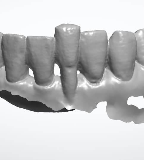
Why this site is so demanding
Lower anteriors are among the most challenging implant sites in the mouth.
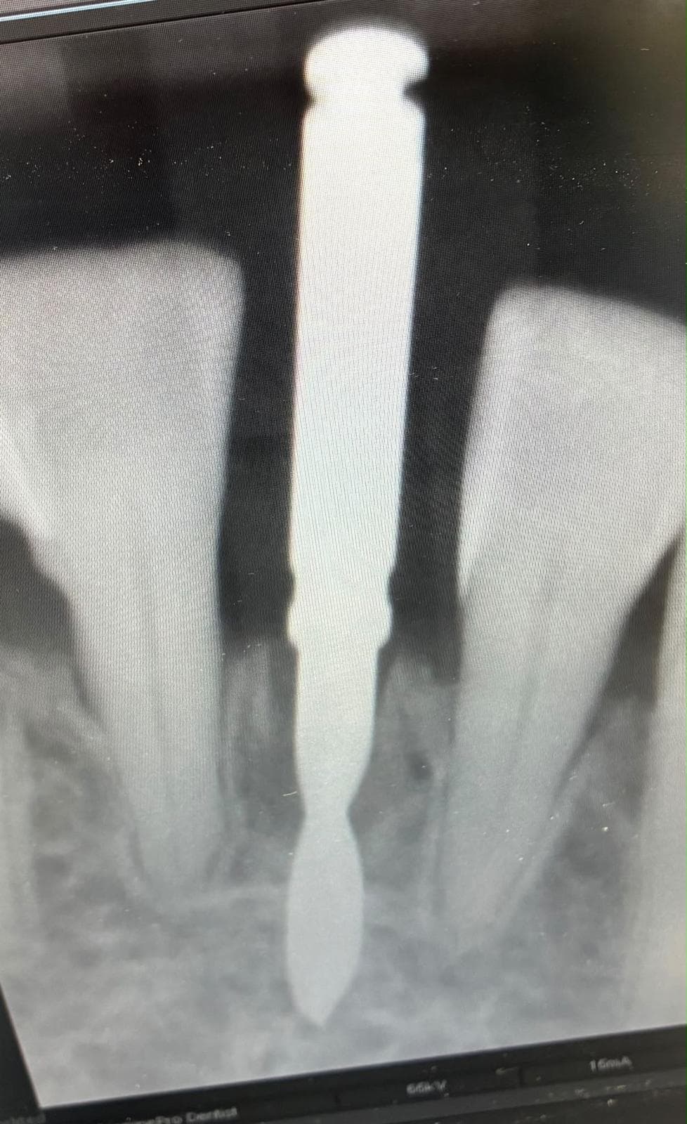
- Very little space: They are the narrowest teeth, so there’s little mesiodistal space to work with. The established guidance is a minimum of 1.5 mm between implant and adjacent root. That distance protects the interproximal bone, the blood supply and the vitality of the neighbouring teeth. In a space this narrow, there’s barely any margin either side.
- Immediate placement adds complexity: The implant has to engage the remaining bone of a fresh extraction socket, in the right position within the socket, not simply following the path of the root that was removed.
- The tooth itself can’t be used as a guide: Because the LL1 was over-erupted, the new crown needs to sit back in line with the arch, not where the old tooth was. The implant must be planned for where the restoration should be, not where the tooth happened to be.
- Recession raises the stakes: With soft tissue already compromised, the implant’s position and angulation have a direct effect on how well the gingiva is supported around the final crown.
With so little margin, freehand placement leaves almost no room for error. A small deviation in angle or depth can bring the implant too close to an adjacent root, or leave the crown with a poor emergence profile.
How digital planning solves it
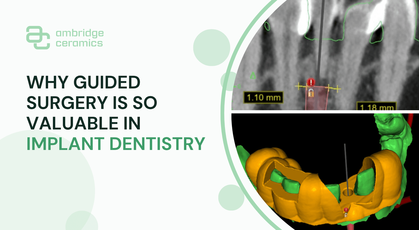
Digital planning combines the CBCT scan, which shows the bone and roots, with the intraoral scan, which shows the teeth and soft tissue. Together they give a complete 3D picture of the site.
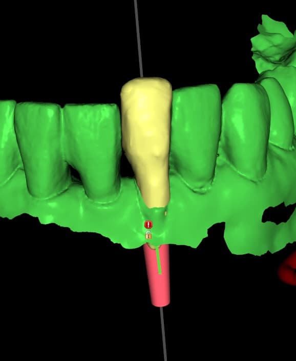
From there, the final restoration is designed first, and the implant is planned around it. Its position, angulation and depth are set to support the ideal crown, and the distance to each adjacent root is measured and confirmed before the patient is even in the chair.
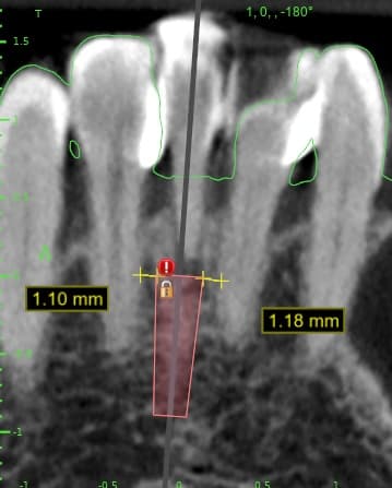
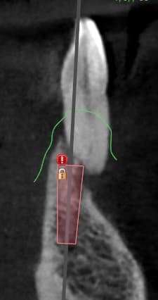
The plan is then turned into a 3D printed surgical guide that fits precisely over the teeth. During surgery, the guide controls the position, angle and depth of each drill, so the implant goes exactly where it was planned.
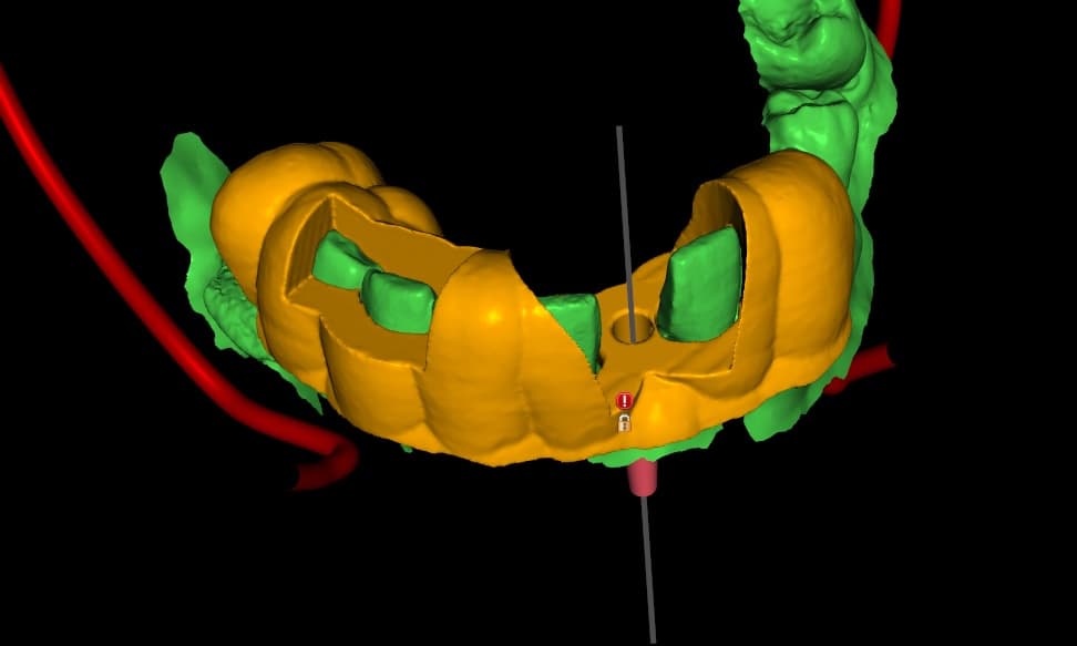
We designed and printed the surgical guide here at Ambridge Ceramics, working from the implant clinician’s CBCT and intraoral scan, so every measurement was confirmed before the guide left the laboratory.
The benefits
Accuracy
The planned position is transferred directly to the patient.
Safety
Distances to adjacent roots and other structures are confirmed before surgery, not estimated during it.
Restoration-led results
The implant supports the ideal crown, with a better emergence profile and soft tissue support.
Efficiency
Surgery is often quicker and more predictable, which means a more comfortable experience for the patient.
The result
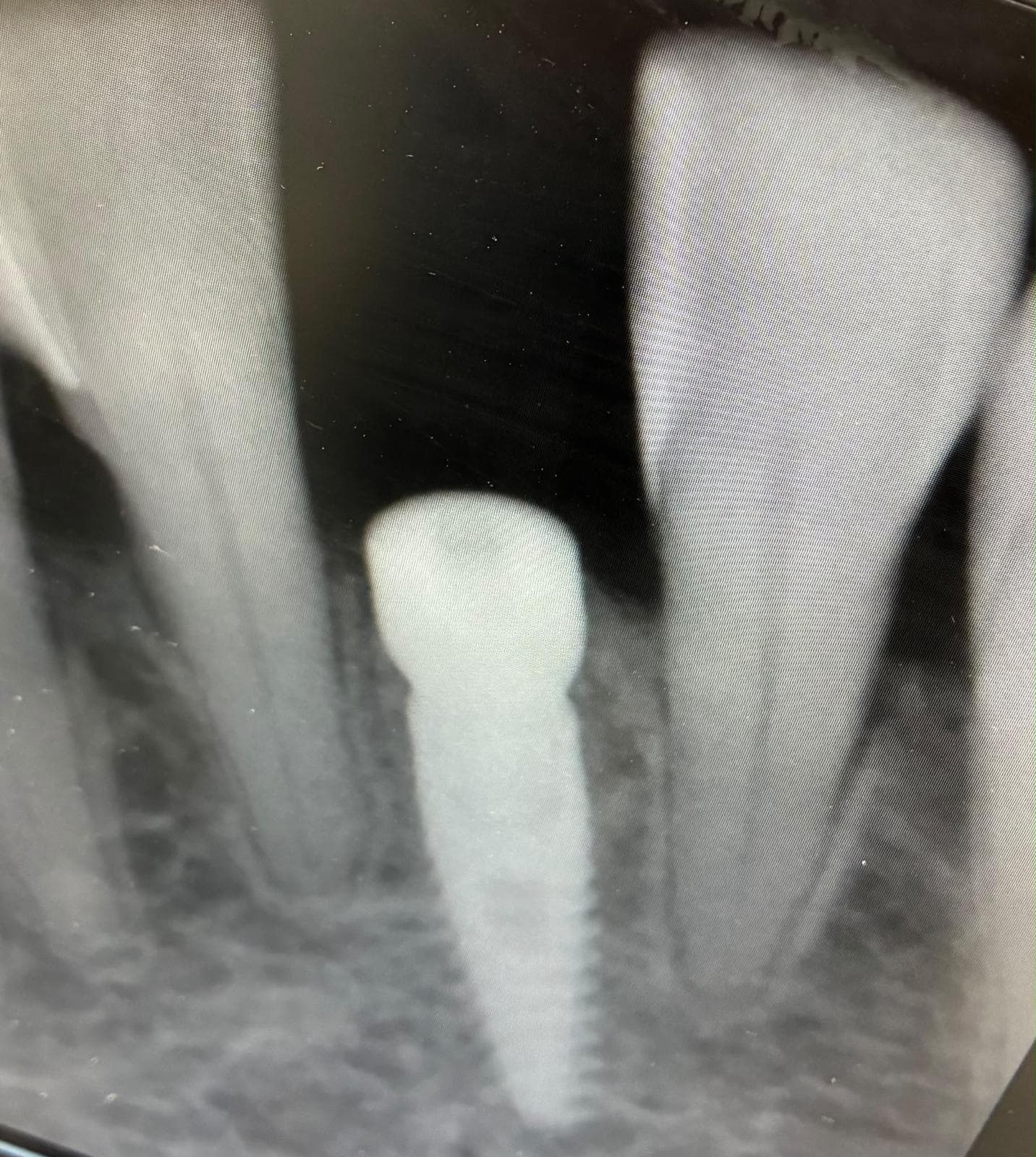
In our client’s words: “We managed to squeeze this one in nicely.”
It’s a modest way of describing a technically demanding placement. It also shows what guided surgery makes possible: even the narrowest sites can be treated with confidence.
Planned precisely, placed predictably, and restored with confidence.
Have a demanding implant site coming up?
Send us your CBCT and intraoral scan and we’ll plan and print a surgical guide for your case.
Ready to Work With Ambridge Ceramics?
Talk to our dental laboratory team about your restoration requirements and laboratory workflow. Experience consistent quality, digital precision, and technician-led technical support for your implant cases.